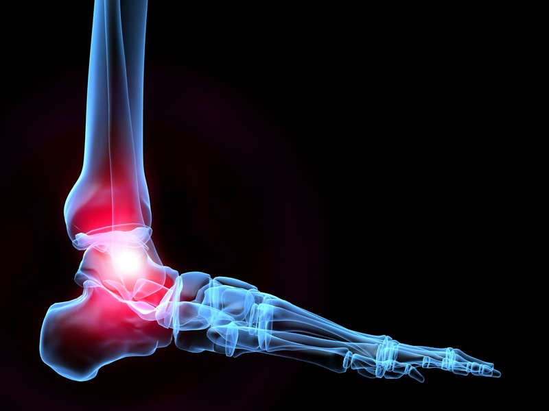
The foot and ankle are hugely complex…
Hence why the profession of chiropody, now more commonly known as Podiatry has been around in one format or another for hundreds of years.
The images I have displayed to coincide with this blog post are to show just how useful ultrasound is as an imaging technique.
X-ray is used as a mainstay imaging modality by GP’s to ‘see what’s going on’, an X-ray can be hugely beneficial to look within the joints, to see arthritis, signs of gout and fractures to name just a few. But, what X-ray isn’t very good at is looking at swelling.
This is where ultrasound really comes into its own!
Ultrasound works well as your Podiatrist can easily and quickly compare one foot to the other, which helps with diagnosis.
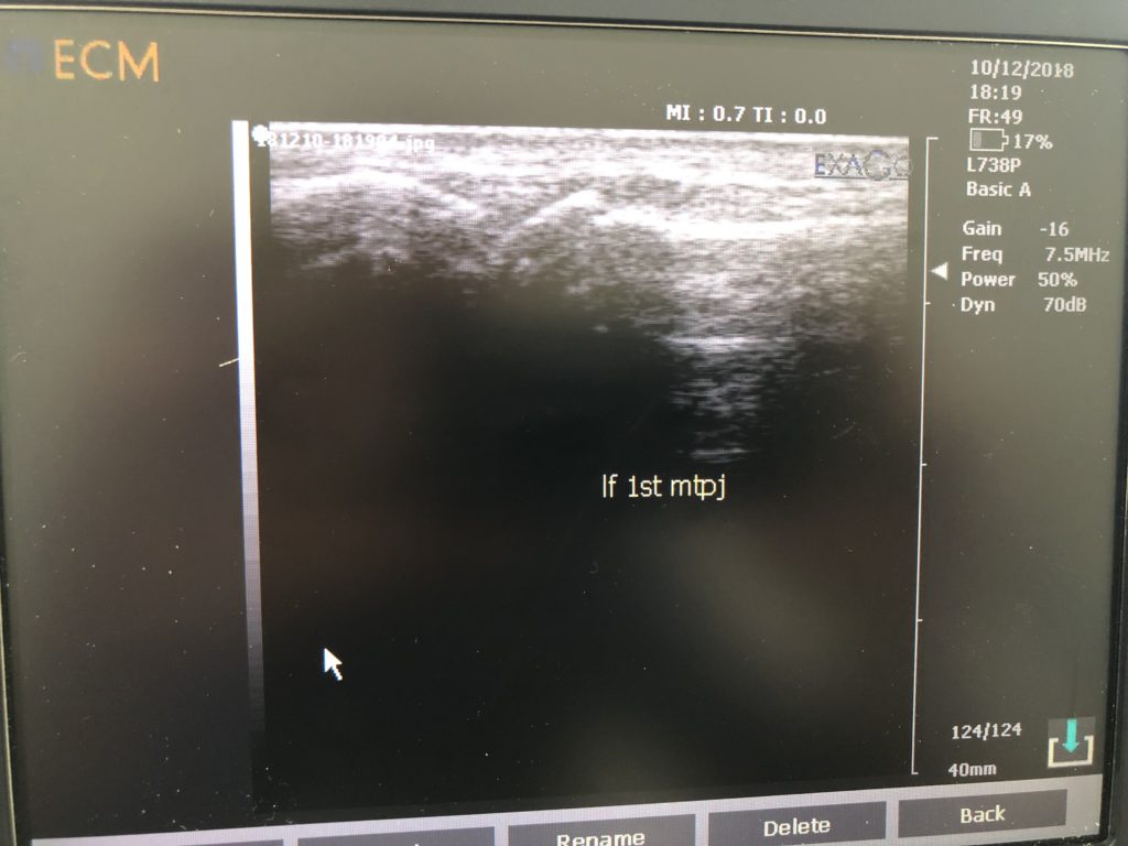
The images below show how a patient’s left big toe joint is normal, but the right foot big toe joint isn’t. This was where the pain was coming from when the patient arrived at the clinic following a trip up the stairs.
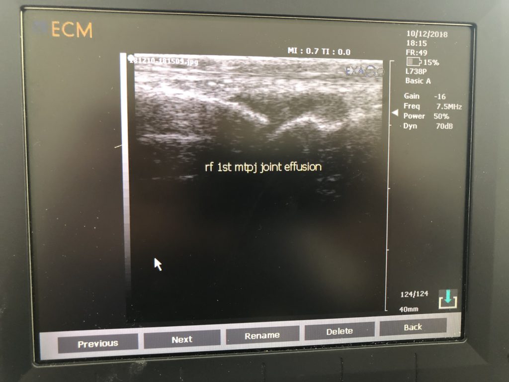
The reference to the word ‘effusion’ within the image means swelling and all the black within the groove is essentially swollen fluid that has collected as a result of trauma. What is interesting to mention with this case is there is no arthritis associated with the joint, it’s perfectly healthy, apart from the injury described.
Ultrasound was able to show the patient just how swollen their toe was and why it was feeling so sore when it was bent. The swelling made for a hugely stiff joint. This was settled down with an insole to stop the joint being used and took away 60% of the pain, with a steroid injection used thereafter to remove the rest. The patient was back to running in no time!
And remember… always be careful on stairs!
If you have any questions or would like to book an appointment, get in touch with us on 01444 453874 or on our contact page!
